
The technique.
The staining.
Similar assays.
The technique
“Immuno” refers to immunology. Fluorescence refers to the phenomenon of light absorption and its emission, typically in the ultraviolet or visible spectrum. For example, some chemicals will absorb blue light (around 450nm) emit green light (around 495nm).
Antibodies are proteins secreted by immune cells (B lymphocytes/ B cells) against foreign proteins (antigens). So, antigen= antibody generator.
Ideally, any protein can induce the secretion of antibodies. Human proteins can induce mouse B cells to secrete antibodies. In this case, we say that the antibody is “raised in mouse.”
Thus, the primary antibody will be used against any protein one wishes to identify. Then, a secondary antibody, an “anti-antibody”, is used to identify the primary antibody. The secondary antibody contains a fluorophore or the chemical compound that emits light.
Finally, one can observe this fluorescent light under the microscope.
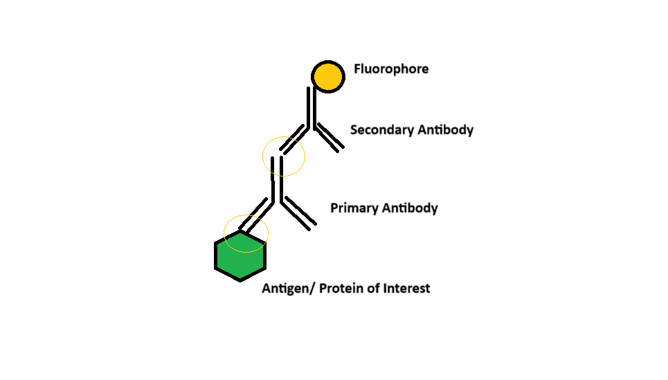
Disclaimer: The coloring provided here is just for representation and are not the actual coloring of these proteins and chemicals.
For the selection of the secondary antibody, one has to know what the primary antibody was “raised in.” For example, if the primary antibody was raised in mouse, the secondary will be raised in another “host” animal, say donkey.
Why? Because the anti-antibody is formed against the primary antibody. In this case, the primary antibody acts as foreign protein against which the B cells of the “host” react.
Lastly, the fluorescence.
Fluorescein is a chemical that is used clinically. A fluorescein injection has been used to understand certain parts of the eye since the contrasting colors can help identify any corneal or vessel abnormalities.
Similarly, a fluoroscent dye or molecule attached to antibodies can specifically help to identify the location of particular antigens bound to the antibody. In the laboratory, we use Alexa Fluor 546 and 488.
In the spectrum, Alexa Fluor 546 is similar to a red dye, like phalloidin used to visualize actin in the cytoskeleton. These are identified (or the antibody is “labelled”) by tetramethylrhodamine isothiocyanate (TRITC).
The staining
Immunofluorescence is used to visualize tissues or cells under a microscope. Since antigen-antibody reactions are very specific, this technique is specific for the protein of interest.
A general protocol is to “fix” the cells/ tissue sections on a glass slide. 4% paraformaldehyde (PFA) is used for this. Fixing is important to retain the structure of the tissues and halt the decomposition process. Once fixed, the “blocking” is performed to prevent the antibody binding to other random proteins which may be extremely similar to the protein of interest. I mentioned it previously when I was writing about identifying proteins. Then we can start with addition of the antibodies and finally, visualizing.
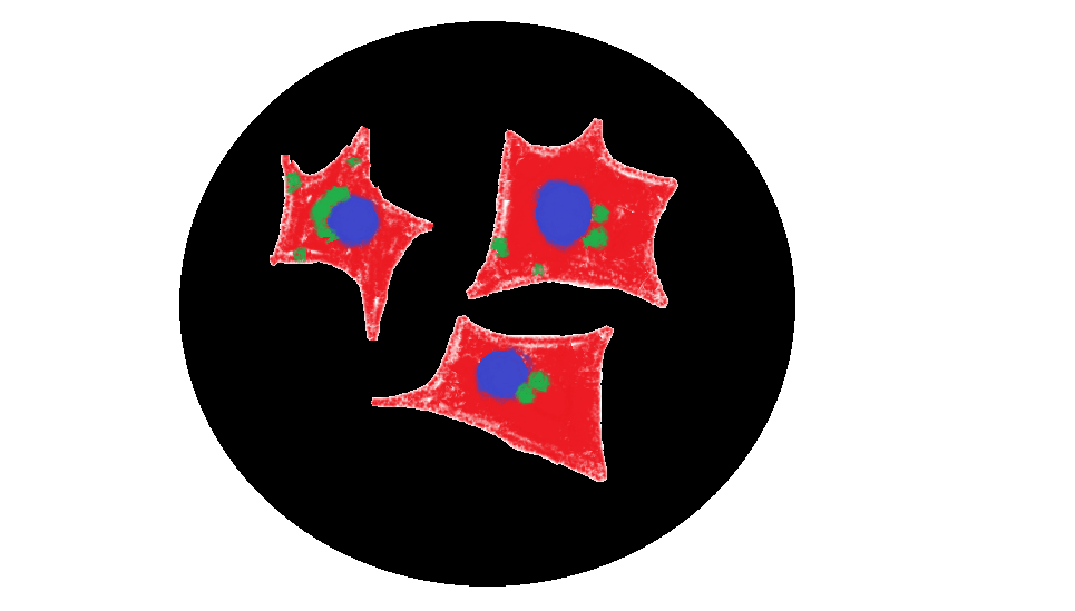
Personally, I enjoy the result of this assay. The cells appear very pretty under the confocal microscope and I would urge the reader to Google some IF images of cells or tissues.
Similar Assays
An assay is an analytical procedure to detect or quantify or study any (biological) entity such as a protein.
Western blot is a technique that uses a similar immunological principle to identify proteins.
ELISA or Enzyme-linked immunosorbent assay is another technique which uses the same principle to identify proteins (or antigens). Most pregnancy kits, viral diagnostic kits such as for dengue, hepatitis or HIV use this technique.


Leave a comment